


ELBOW
Ioannis Raftakis
The ultrasound of the elbow is a useful complementary method for the evaluation of elbow pathological conditions, allowing detection of changes that are undetectable by clinical examination and plain x-rays.
Advantages of the method are:
Highlighting the presence of fluid in the elbow joint (figure 1a), in all or within individual recesses – Coronoid (figure 1b), or Annular recess (figure 1c), while the small amounts of fluid can’t be clinically detected.
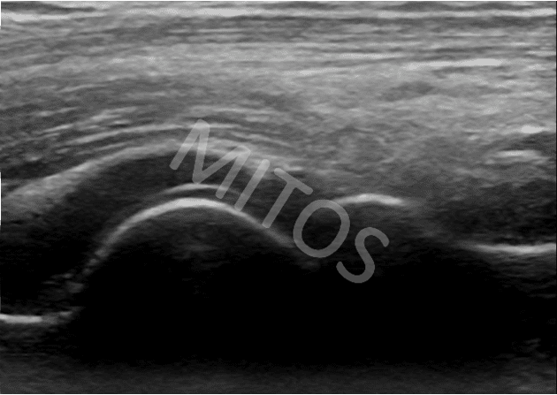
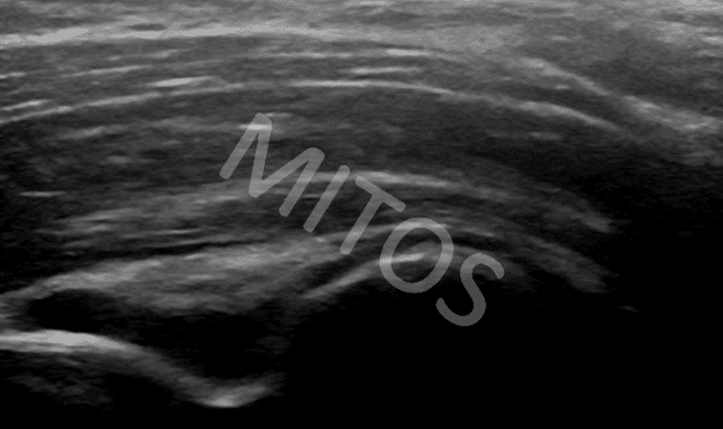
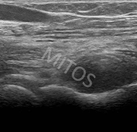
Ultrasonographic examination easily depicts the synovial hypertrophy, (figure 1d).
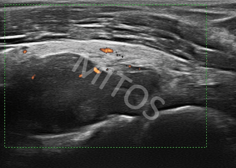
Doppler mode can highlight the presence of inflammation.
Tendon lesions
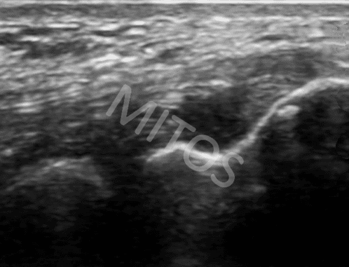
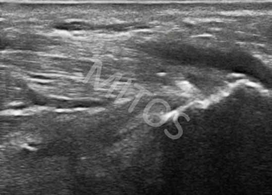
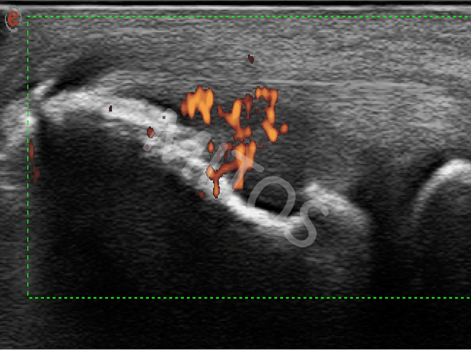
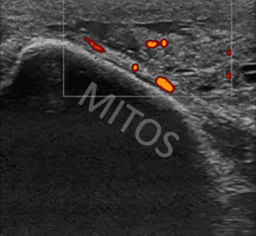
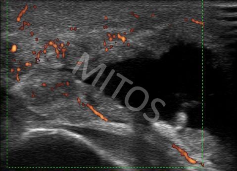
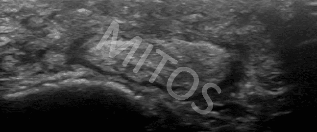
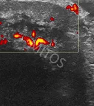
Ultrasound can depict acute inflammation – acute lateral epicondylitis – (figure 2a), or acute medial epicondylitis (figure 2b), as well as their chronic degenerative changes or the presence of tears. The use of Doppler mode can highlight the presence of inflammation within the tendinous structures, (figure 2c).
Imaging of olecranon bursitis
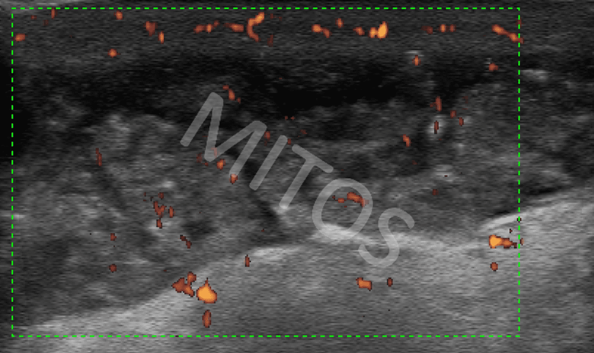
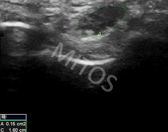
The olecranon bursa can be visualized in a view of synovial hypertrophy or / and the presence of fluid within the cavity in the context of infection (e.g. Staphylococcal infection, (figure 3a, figure 3b)), or inflammatory diseases, (e.g. gout, (figure 3c)).
Nerve disorders
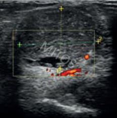
The area of the elbow pass three nerves: ulnar, median and radial nerve. Their possible compression can cause severe symptoms. Ultrasound of these nerves allows their quick and safe evaluation. (e.g. ulnar nerve neuropathy, (figure 4)).
Ultrasound can highlight the presence of free intra-articular bodies and even differentiate rheumatic nodules, (figure 5), from gouty tophi, (figure 6a, figure 6b).
Additionally to imaging, ultrasound also allows US guided punctures by aspiration of even a small amount of synovial fluid aiming at establishing of diagnosis. US guided administration of therapeutic drags locally at the exact point of inflammation is also approved.
Evaluation of therapeutic effect
Ultrasound method can be used in monitoring of the therapeutic response in treated patients with systemic diseases or of patients to whom intra-articular injection had been applied during the disease course.
Bibliography:
- Backhaus M, Burmester GR, Gerber T etal: Guidelines for musculoskeletal ultrasound in rheumatology. Ann Rheum Dis. 60(7), 641-649 (2001).
- Kahle W, Leonhardt H, Platzer W: Taschnatlas der Anatomie 1985.
- Bruyn A.W, Schmidt: Introductory Guide to Musculoskeletal Ultrasound for the Rheumatologist 45-56 2006.
- Bianchi S, Martnoli C: Ultrasound of the Muskuloskeletal System 349-407 2007.
- Nalbant A, Corominas H, Hsu B et al: Ultrasonography for assessment of subcutaneous nodules. J Rheum 2003, 30: 1191-1195.